


.webp)
Pet Ultrasound Scans
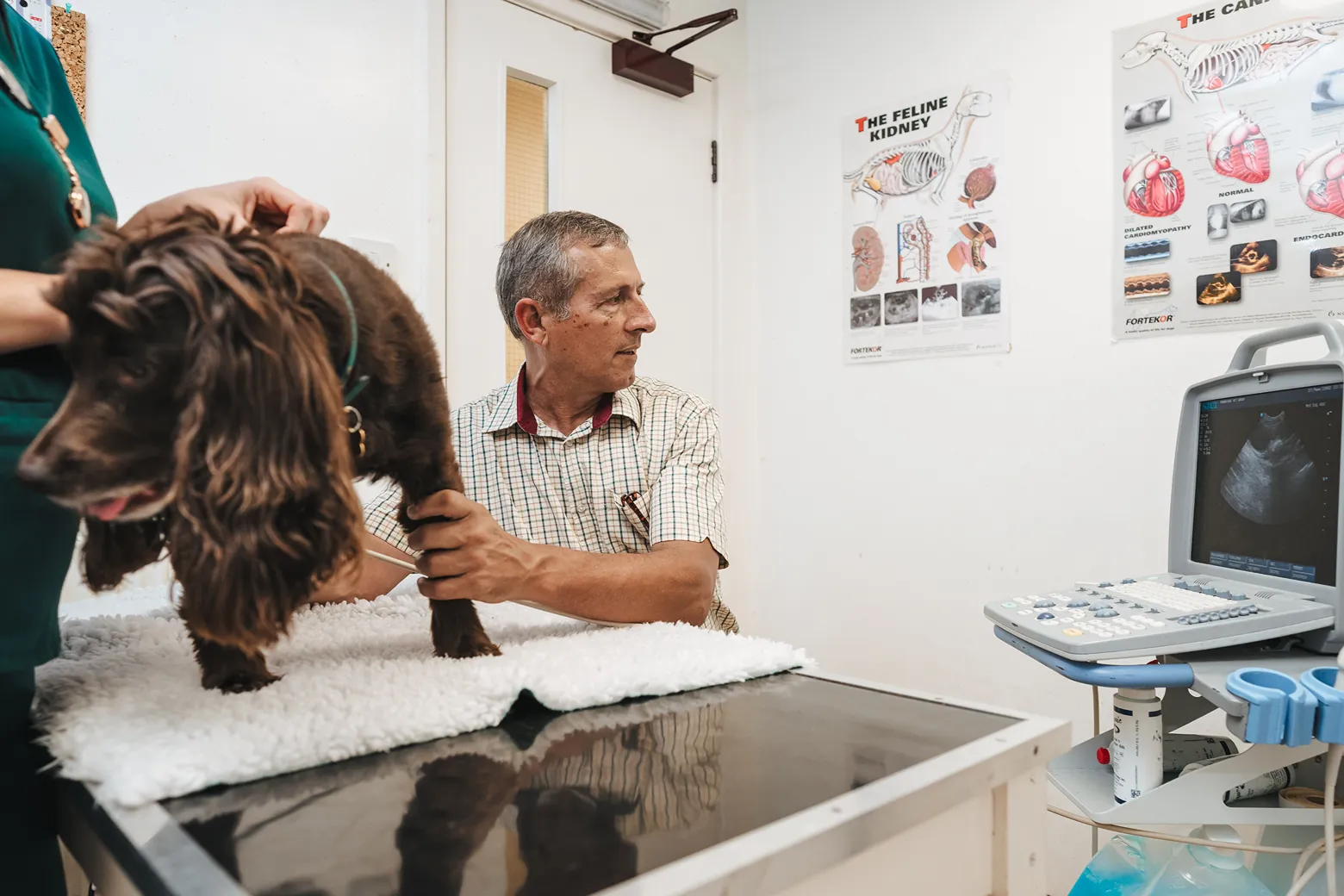
Ultrasound is a safe, painless way to examine your pet's internal organs in real time. It provides detailed information without surgery, allowing our vets to diagnose problems quickly and discuss the results with you during your appointment.
What Can Ultrasound Help Us Diagnose?
Ultrasound is a versatile diagnostic tool and the range of conditions it can help us investigate is broad. For example:
Pregnancy confirmation
As with humans, ultrasound is the most reliable method for confirming pregnancy in your pets. It can detect a pregnancy from around four weeks after mating and, as the pregnancy progresses, allows us to assess foetal development and estimate the number of offspring.
Abdominal organs
The liver, kidneys, spleen, bladder, pancreas and gastrointestinal tract can all be examined in detail using ultrasound. We can assess organ size and texture, identify masses or cysts, detect fluid accumulation or internal bleeding and look for signs of inflammation or structural change that would not be visible on an X-ray.
For a pet that is losing weight without explanation, drinking more than usual, vomiting regularly or has an abnormality detected on physical examination, an abdominal ultrasound is often one of the first investigations we recommend. It complements blood test findings and frequently helps us narrow down a diagnosis considerably.
The heart
An ultrasound examination of the heart (known as an echocardiogram) allows us to assess its structure and function in real time. We can evaluate the size of the chambers, observe how the valves are moving, look for fluid around the heart and assess the direction and speed of blood flow.
This is particularly valuable in any pet where a murmur has been detected during a routine examination. Identifying heart disease early gives us the best opportunity to start treatment at the point where it is most likely to make a long-term difference.
Guided biopsy and sampling
Where we need a tissue or fluid sample from an internal organ or suspected mass, ultrasound allows us to guide a needle to exactly the right location with precision and safety. This improves the quality of the sample obtained and reduces the risk to surrounding structures, making it a much more reliable approach than sampling without direct visualisation.
Emergency assessment
In urgent situations, a focused ultrasound examination can quickly identify internal bleeding, fluid around the lungs or heart or signs of organ rupture. This kind of rapid assessment can be critical in guiding immediate treatment decisions when time matters.
So, you see, the common thread across all of these applications is the ability to see what is happening inside your pet in real time, without surgery or (in most cases) sedation. That means faster answers, less stress for your animal and a clearer path to the right treatment.
Ultrasound and X-Rays: Understanding the Difference
The two imaging methods complement each other rather than compete. X-rays excel at showing bones, joints and chest conditions, where dense structures create clear contrast. Ultrasound, on the other hand, provides far superior detail of soft tissues and abdominal organs.
In many diagnostic workups we use both. An X-ray gives us the broad structural picture; ultrasound fills in the soft tissue detail. Knowing which to use and when is part of what we bring to any diagnostic assessment.
What to Expect on the Day
When you arrive, we take a brief history and carry out a clinical examination before the scan begins. In most cases, no sedation is necessary. For pets that are particularly anxious or in significant discomfort, a mild sedative can help them relax and make the images clearer.
A small area of fur will be clipped over the area being scanned and a gel applied to the skin to ensure good contact with the probe. Your pet will usually be settled comfortably on a padded surface for the duration. Most pets relax into the procedure quickly, without any stress.
Because ultrasound uses sound rather than radiation, it is safe to use repeatedly without any cumulative risk to your pet. Images can be captured as stills or short video clips, which can be stored as part of your pet’s clinical record and allow us to compare findings over time.
Why Choose Bowbridge Vets For Diagnostics?
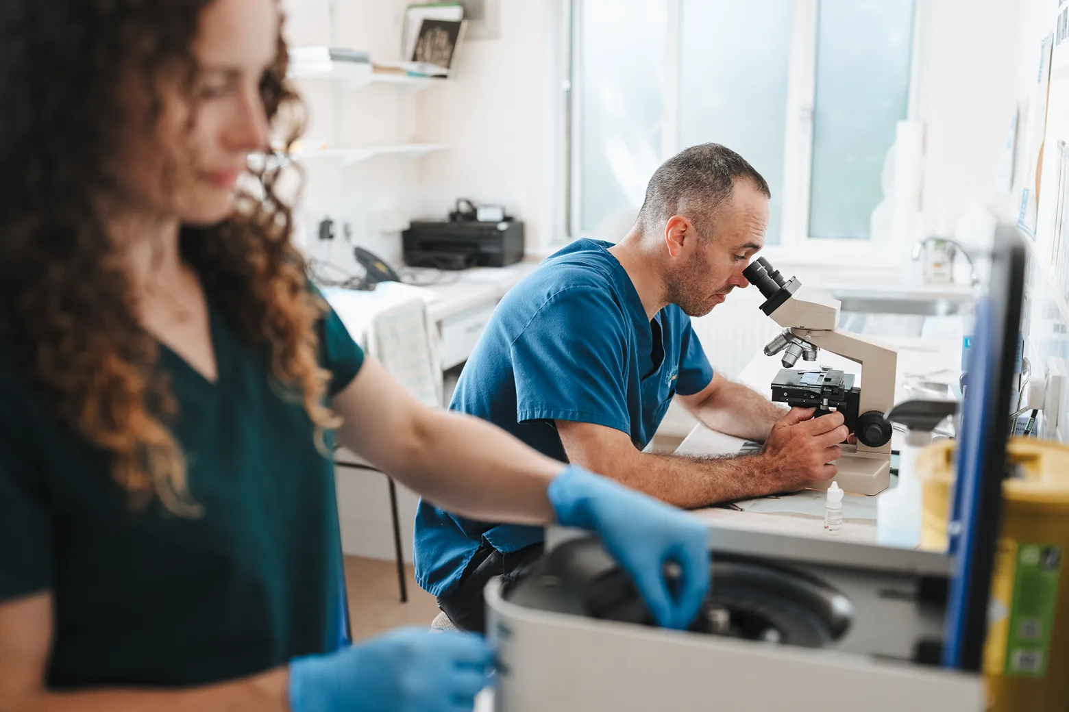
Getting to an answer quickly and accurately matters, especially when your pet is unwell. Our in-house diagnostic equipment means we can run blood tests, take X-rays and carry out ultrasound scans without sending you elsewhere. Also, results are usually available on the same day.
We explain what we find in plain English, before we discuss next steps. We recommend what is clinically appropriate for your pet’s individual situation. When we recommend a test, you will always know exactly why.
We have been caring for the pets of Stroud and Stonehouse since 1967. That experience informs not just how we practise clinically, but how we listen, how we communicate and how we think about the pets in our care.
Frequently Asked Questions
How does veterinary ultrasound work?
The probe emits high-frequency sound waves that pass into the body and bounce back from internal structures at different rates depending on tissue density. These returning signals are translated into real-time images on a monitor, creating a moving picture of the organs and tissues beneath the skin.
What is the difference between ultrasound and X-ray?
X-rays are better for bones, joints and chest conditions. Ultrasound is better for soft tissues and abdominal organs. Both have important roles in veterinary diagnostics and are often used alongside each other rather than as alternatives.
Does ultrasound hurt my pet?
No. Ultrasound is entirely painless. The probe sits gently against the surface of the skin and there is no sensation involved in the sound waves passing through the body. The coolness of the gel and any clipping of fur beforehand are the most noticeable parts of the experience for most pets.
Does my pet need to be sedated for an ultrasound?
In most cases, no. The majority of pets tolerate ultrasound well without sedation, particularly when they are relaxed and handled calmly. Where a pet is very anxious, in discomfort or finding a position difficult to hold, a mild sedative makes the experience more manageable and the images clearer.
Do I need to prepare my pet before an ultrasound scan?
For abdominal scans, we ask that your pet fasts for several hours beforehand. Food in the stomach and gas in the intestines can affect image quality. Water is usually fine up until the time of the appointment. We will give you specific instructions before your appointment.
Will the fur need to be clipped?
Yes, over the area being scanned. The amount clipped is usually small and the fur grows back within a few weeks. Clipping is necessary to ensure good contact between the probe and the skin, which directly affects the quality of the images we can produce.
Can ultrasound detect cancer in pets?
Ultrasound can identify masses, abnormal organ texture and other changes that may indicate cancer. It cannot provide a definitive diagnosis on its own: for that, a biopsy and laboratory analysis are usually needed. Ultrasound is often the starting point for investigating a suspected mass and, crucially, can be used to guide the biopsy needle to exactly the right location.
How soon will I get the results?
Scan findings are discussed with you on the day. If tissue samples have been taken for analysis, laboratory results typically return within five to seven working days. Where images are sent for specialist radiologist review, a written report usually comes back within a few days. We contact you as soon as results are available.